

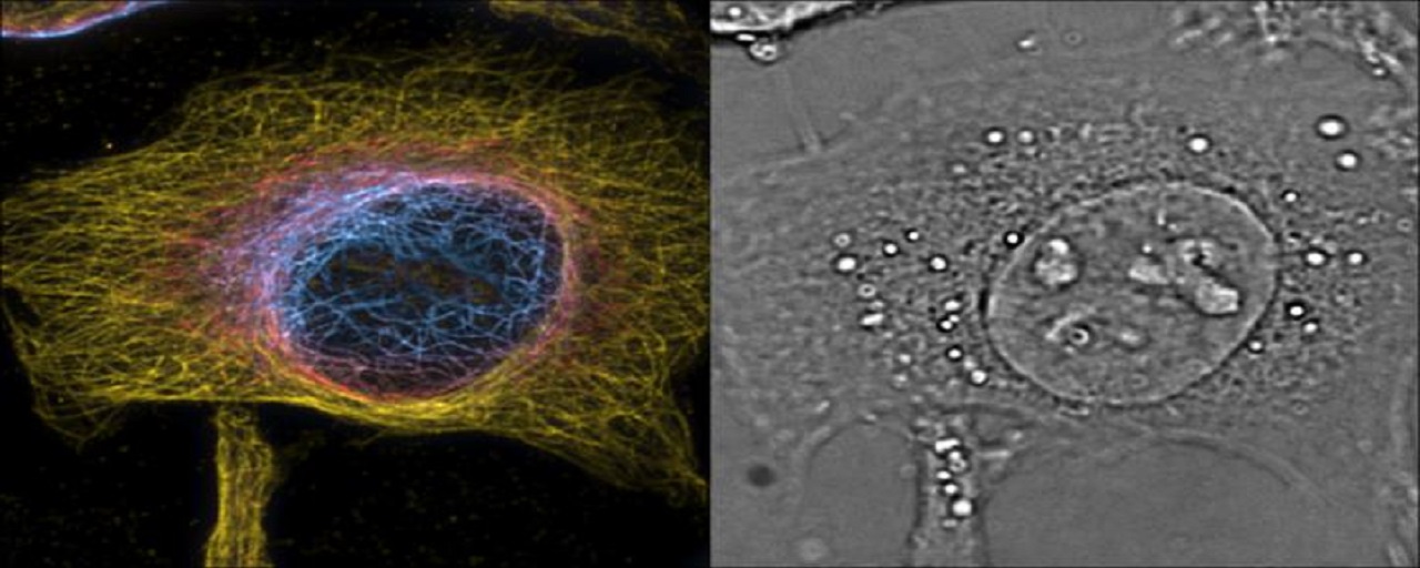
Super-resolution microscopy has been known to have the ability to capture a high-quality view of interior structures and organelles in cells. However, it is severely restricted, because this only works if examining something that remains static. Living cells would require a much more complex way to be able to study them as thoroughly. Luckily, a team led by Professor Theo Lasser, the head of the Laboratory of Biomedical Optics (LOB) have developed a way to do this.
Super-resolution microscopy has been known to have the ability to capture a high-quality view of interior structures and organelles in cells. However, it is severely restricted, because this only works if examining something that remains static. Living cells would require a much more complex way to be able to study them as thoroughly. Luckily, a team led by Professor Theo Lasser, the head of the Laboratory of Biomedical Optics (LOB) have developed a way to do this. They have combined the sensitivity and high time-resolution of phase imaging with the specificity and high spatial resolution of fluorescence microscopy in order to visualize cells in 3D. To read more, check out thefull article.
Filed Under:Rapid prototyping


